No one single laboratory test is 100% accurate in diagnosing all types of thyroid disease; however, a combination of two or more tests can usually detect even the slightest abnormality of thyroid function. For example, a low T4 level could mean a diseased thyroid gland or a non-functioning pituitary gland not stimulating the thyroid function. Since the pituitary gland would normally release TSH if the T4 is low, a high TSH level is also present; this confirms that the thyroid gland (not the pituitary gland) is responsible for the hypothyroidism. If the T4 level is low and TSH is not elevated, the pituitary gland is more likely to be the cause for the hypothyroidism. Of course, this would drastically effect the treatment since the pituitary gland also regulates the body's other glands (adrenals, ovaries, and testicles) as well as controlling growth in children and kidney function. Pituitary gland failure means that the other glands may also be failing and other treatment than just thyroid may be necessary. The most common cause for the pituitary gland failure is a tumor of the pituitary and this might also require surgery to remove. The older tests of thyroid function: Basal Metabolic Rate (BMR), basal body temperature, Achilles Heel Reflex Time, and measurement of Protein Bound Iodine (PBI) have been replaced by much more accurate thyroid function tests. Modern measurement of thyroid hormones is done by a new technique, radioimmunoassay (RIA), discovered by Dr. Solomon A. Berson and Dr. Rosalyn S. Yallow. They were awarded the 1977 Nobel Prize in Medicine for this discovery which revolutionized the study of thyroid disease as well as the entire field of endocrinology. The following tests of the thyroid are those currently used today.
A. Measurement of Thyroid HormonesT4 by RIA (radioimmunoassay) is the most used thyroid test; it is frequently referred to as a T7 which means that a resin T3 uptake (RT3u) has been done to correct for certain medications such as birth control pills, other hormones, seizure medication, cardiac drugs, or even aspirin that may alter the routine T4 test. The T4 (or T7) reflects the amount of thyroxine in the blood. If the patient does not take any type of thyroid medication, this test is usually a good measure of thyroid function. As stated earlier, thyroxine represents 80% of the thyroid hormone produced by the normal gland and generally represents the overall function of the gland. The other 20% is triiodothyronine measured as T3 by RIA. Sometimes the diseased thyroid gland will start producing very high levels of T3 but still produce normal levels of T4. Therefore measurement of both hormones provides an even more accurate evaluation of thyroid function.Most of the thyroid hormones in the blood are attached to a protein called thyroid binding globulin (TBG). If there is an excess or deficiency of this protein it alters the T4 or T3 measurement but does not affect the action of the hormone. If a patient appears to have normal thyroid function, but an unexplained high or low T4, or T3, it may be due to an increase or decrease of TBG. Direct measurement of TBG can be done and will explain the abnormal value. Excess TBG or low levels of TBG are found in some families as an hereditary trait. It causes no problem except falsely elevating or lowering the T4 level. These people are frequently misdiagnosed as being hyperthyroid or hypothyroid, but they have no thyroid problem and need no treatment.
B. Measurement of Pituitary FunctionTSH by IRMA (immunoradiometric assay) is produced by the pituitary gland. Normally, low levels (less than 5 units) of TSH are sufficient to keep the normal thyroid gland functioning properly. When the thyroid gland becomes inefficient such as in early hypothyroidism, the TSH becomes elevated even though the T4 and T3 may still be within the "normal" range. This rise in TSH represents the pituitary gland's response to a drop in circulating thyroid hormone; it is usually the first indication of thyroid gland failure. Since TSH is normally low when the thyroid gland functions properly, the failure of TSH to rise when circulating thyroid hormones are low is an indication of impaired pituitary function. The new "sensitive" TSH test will show very low levels of TSH when the thyroid is overactive. Interpretations of the TSH level depends upon the level of thyroid hormone; therefore, the TSH is usually used in combination with other thyroid tests such as the T4 RIA and T3 RIA.TRH test: In normal people TSH can be increased by giving TSH Releasing Hormone (TRH). A baseline TSH of 5 or less usually goes up to 10-20 after giving an injection of TRH. Patients with too much thyroid hormone (thyroxine or triiodothyronine) will not show a rise in TSH when given TRH. This "TRH test" is presently the most sensitive test in detecting early hyperthyroidism. Patients who show too much response to TRH (TSH rises greater than 40) may be hypothyroid. This test is also used in cancer patients who are taking thyroid replacement to see if they are on sufficient medication. It is sometimes used to measure if the pituitary gland is functioning. The new "sensitive" TSH test has eliminated the necessity of performing a TRH test in most clinical situations.
C. Iodine UptakeA third parameter of measuring thyroid function is to measure how much iodine is taken up by the thyroid gland (RAI uptake). Hypothyroid patients usually take up too little iodine and hyperthyroid patients take up too much iodine.The test is performed by giving a tracer dose of radioactive iodine on an empty stomach. The iodine is concentrated in the thyroid gland or excreted in the urine over the next few hours. The amount of iodine that goes into the thyroid gland can be measured by a "Thyroid Uptake". Of course, patients who are taking thyroid medication will not take up iodine in their thyroid gland because their own thyroid gland is turned off and is not functioning. At other times the gland will concentrate iodine normally but will be unable to convert the iodine into thyroid hormone; therefore, interpretation of the iodine uptake is usually done in conjunction with blood tests.
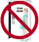
D. Thyroid ScanTaking a picture of the thyroid gland requires giving a radioisotope to the patient and letting the thyroid gland concentrate the isotope. Therefore, it is usually done at the same time that the iodine uptake test is performed. Although other isotopes, such as technetium, will be concentrated by the thyroid gland; these isotopes will not measure iodine uptake. It has also been found that thyroid nodules that concentrate iodine are never cancerous; this is not true if the scan is done with technetium. Therefore, all scans we do are now done with iodine. Pregnant women should not have thyroid scans performed.
Two types of thyroid scans are available: A rectilinear scan draws a picture of the gland to scale on paper. This is the only type of scan that can be used to measure the size of the gland. It is also best for identifying the location of nodules when needle biopsies are necessary. This type of scan is obtained by the scanner moving back and forth over the gland and takes approximately forty-five minutes to perform.
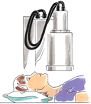
Rectiliniear Thyroid Scan A camera scan is done using a gamma camera. The same camera as is used for scanning the liver, brain, etc. The camera operates in a fixed position and views the entire thyroid gland at once. This type of scan takes only five to ten minutes, but the image produced is not related to the actual size of the thyroid gland, and the gland looks distorted similar to a picture taken with a camera using a fisheye lens.
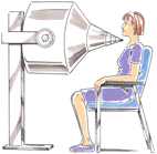
Camera Thyroid Scan A rectilinear scan is the preferred method of scanning. In the 1990's, a new scanner called a Computerized Rectilinear Thyroid (CRT) scanner was introduced. The CRT scanner utilizes computer technology to improve the clarity of thyroid scans and enhance thyroid nodules. It measures both thyroid function and thyroid size. A lifesize 1:1 color scan of the thyroid is obtained giving the size in square centimeters and the weight in grams. The precise size and activity of nodules in relation to the rest of the gland is also measured.
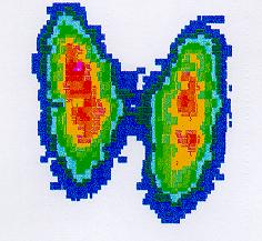
CTS of the normal thyroid gland
In addition to making thyroid diagnosis more accurate, the CRT scanner improves the results of thyroid biopsy. The accurate sizing of the thyroid gland aids in the follow-up of nodules to see if they are growing or getting smaller in size. Knowing the weight of the thyroid gland allows more accurate radioactive treatment in patients who have Graves' disease. Thyroid scans are used for:
E. Thyroid UltrasoundThyroid ultrasound refers to the use of high frequency sound waves to obtain an image of the thyroid gland and identify nodules. It tells if a nodule is "solid" or a fluid-filled cyst, but it will not tell if a nodule is benign or malignant. Ultrasound allows accurate measurement of a nodule's size and can determine if a nodule is getting smaller or is growing larger during treatment.
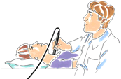
Ultrasound measurement Ultrasound aids in performing thyroid needle biopsy by improving accuracy. More recently, ultrasound has become a primary way to follow patients who have been operated on for thyroid cancer to detect early recurrence.
F. Thyroid AntibodiesThe body normally produces antibodies to foreign substances such as bacteria; however, some people are found to have antibodies against their own thyroid tissue. A condition known as Hashimoto's Thyroiditis is associated with a high level of these thyroid antibodies in the blood. Whether the antibodies cause the disease or whether the disease causes the antibodies is not known; however, the finding of a high level of thyroid antibodies is strong evidence of this disease. Occasionally, low levels of thyroid antibodies are found with other types of thyroid disease. The presence of thyroid antibodies has recently been associated with an increase frequency of miscarriage. When Hashimoto's thyroiditis presents as a thyroid nodule rather than a diffuse goiter, the thyroid antibodies may not be present.
G. Thyroid Needle BiopsyThis has become the most reliable test to differentiate the "cold" nodule that is cancer from the "cold" nodule that is benign ("hot" nodules are never cancerous). It provides information that no other thyroid test will provide. While not perfect, it will provide valuable information in 75% of the nodules biopsied. The needle biopsy has reduced the number of patients who are operated on for benign nodules and also permitted earlier diagnosis in those patients who have cancer. In the past, when thyroid surgery was performed on a thyroid nodule, only one lobe of the thyroid was removed. If the nodule turned out to be cancerous, the patient was then re-operated upon to remove the rest of the thyroid gland. Today, if the thyroid biopsy shows cancer, the entire thyroid gland is removed with the first operation, therefore avoiding a second unnecessary operation.Thyroid needle biopsy is an office procedure and requires no anesthesia. Except for minor bleeding, no complications have occurred. The purpose of the biopsy is to rule out or confirm malignancy. About 20% of such biopsies are inconclusive and the thyroid pathologist cannot be certain if the tumor is benign or malignant. In such cases, a decision to operate is made upon other criteria. About 5% of biopsies do not yield enough cells to make a diagnosis. In checking a person's thyroid gland, most tests need to be done while not taking thyroid medication. Thyroid antibodies, thyroid ultrasound, and thyroid needle biopsy are not affected by such medicine. The other thyroid tests measure both the thyroid we make and the thyroid we take. These tests will determine if there is too much or too little thyroid in the blood, but do not determine how much comes from the thyroid gland and how much comes from the thyroid pills.
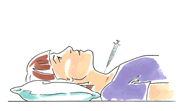
Thyroid Needle Biopsy Occasionally, it is important to measure a person's thyroid status who is on medications such as doing a thyroid scan on a patient who had surgery for thyroid cancer, or a person who was put on thyroid without adequate testing before being started on medication. Since Euthyrox or Synthroid, and most other thyroid pills behave exactly as normal human thyroid hormone, they are not rapidly cleared from the body as other medications are. Most thyroid pills have a half life of 6.7 days which means they must be stopped for five weeks (five half lives) before accurate thyroid testing is possible. An exception to the long half life of thyroid medication is Cytomel - a thyroid pill with a half life of only forty-eight hours. Therefore it is possible to change a person's thyroid replacement to Cytomel for one month to allow time for his regular pills to clear the body. Cytomel is then stopped for ten days (five half lives) and the appropriate test can then be done. Usually patients, even those who have no remaining thyroid function, tolerate being off thyroid replacement only ten days quite well. |


