|
| |
FNAB can provide a specific diagnosis in 70–85% of thyroid nodules. This group comprises the majority
of both benign and malignant lesions. When considering only diagnostic FNAB, sensitivity, specificity
and accuracy are high43. The clinical decision may rely, therefore, mainly on FNAB results. In the
remaining 15–30% of cases FNAB cannot provide a definite diagnosis. In this group, which includes
inadequate samples and “indeterminate” diagnoses, FNAB data should be integrated by additional information
to finalize nodule treatment. Clinical implications of FNAB results will be briefly summarized:
Benign lesions (nodular goiter and thyroiditis)
Nodular goiter and chronic thyroiditis represent the large majority of benign lesions, and account for
approximately 70 % and 5% of all nodules, respectively. Macrofollicular adenomas are often classified
as nodular goiters because of many overlapping aspects.
The real cancer risk in this group is difficult to calculate because many patients are not addressed to
surgery. However, several lines of evidence indicate a cancer risk in the range of 0.5–3 %, which depends
on both the “a priori” cancer risk in a given population and the accuracy reached by the FNAB operator4,25,42.
Given the slow evolution of the majority of differentiated thyroid carcinomas, the cancer mortality due
to this proportion of false negatives is considered very low and clinical follow-up with echographic
measurement of nodule size is appropriate. Evidences that small nodule size and presence of abundant
colloid are predictive for nodule shrinkage after L-thyroxine treatment maybe useful in the decision
whether or not to treat the patient with L-thyroxine43. Thyrodectomy can be reserved for patients
with compression symptoms or esthetic problems. In patients with nodules benign at cytology but with
clinical and/or echographic signs of suspect (e.g. hard consistency, rapid growth, loco-regional lymph
nodes, microcalcifications, etc.) the possibility of surgery should be carefully considered and treatment
individually tailored. In these patients the risk of mortality for the late discovery of a thyroid cancer
should be balanced against the risk of immediate surgery.
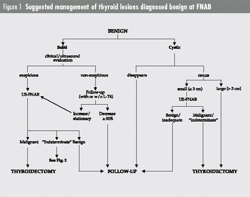
The clinical management of multinodular goiters should be based on FNA results of all palpable nodules.
Particular attention should be paid to cystic/complex nodules because they have a cancer risk similar
to that of solid nodules but a higher false negative rate (up to 17–30%) with higher values observed in
cysts larger than 3 cm45,46. Complete cyst disappearance after aspiration strongly suggests a benign
lesion.
A proposed algorithm for the management of solid or cystic benign lesions is given in Fig. 1.
During the follow-up of benign nodules FNAB repetition is advised by many clinicians in order to
reduce false negatives. The clinical value of rebiopsy is, however, controversial. Some authors favor the
routine use of rebiopsy while others suggest that a repeated FNAB should be reserved to nodules that
enlarge or show other suspicious clinical changes during the follow-up47. It is likely that a second
FNAB can change the initial diagnosis from benign to malignant in 1–2% of cases and is, therefore,
worthwhile48,49. More questionable is the value of a third FNAB, unless the nodule characteristics change
with time.
Malignant lesions (papillary, medullary and undifferentiated
thyroid carcinomas, lymphomas, metastatic nonthyroid cancers).
Papillary cancer is diagnosed in 3–4 % of all nodules while either medullary or undifferentiated cancer is
diagnosed in approximately 1% of nodules. Lymphomas and metastases of non-thyroidal tumors
represent a small minority of asymptomatic thyroid nodules.
In nodules diagnosed malignant at FNAB the cancer risk ranges 97–99%. Therefore, the patient should be
addressed to immediate surgery and the surgeon can plan a total or near total thyroidectomy on the basis
of FNAB data. Intraoperative frozen sections are of little help in these cases50. Serum calcitonin is often
elevated in case of medullary cancer and may be useful to confirm this diagnosis51.
The patient should be informed of the small possibility of a false positive. False positive, however, do
not represent a significant clinical problem since are mostly represented by atypical adenomas that are
best treated by surgery (Table 1).
“Indeterminate” thyroid lesions
Most authors advise thyroidectomy in patients with cytologically “indeterminate” thyroid lesions. However,
these lesions represent a substantial proportion (15–25%) of all thyroid nodules and are malignant
only in 10–20% of cases. It is obvious, therefore, that a refinement of this diagnosis would spare many
unnecessary thyrodectomies. To this aim a variety of approaches have been proposed. Although no consensus
is so far reached, some of these approaches are promising and appear clinically relevant.
Refining diagnosis in “indeterminate” thyroid lesions: subgrouping in classes of
different cancer risk
“Indeterminate” lesions may be easily subgrouped on the basis of simple morphologic or clinical parameters.
This approach, however, is clinically useful only if a subgroup can be identified with a cancer
risk low enough to avoid immediate surgery.
- morphological criteria
The presence of classical features, such as microfollicles, cell atypias, Hurthle cells and clear nuclei may
be used to subgroup lesions with different cancer risk.
Some authors classify nodules in three or four categories: follicular neoplasm, Hurthle cell neoplasm,
suggestive of cancer (papillary or follicular) carcinoma. Afourth category may contain miscellaneous
lesions26. The cancer rate ranges 13–15% in the first category, 14–24% in the second and 50–60% in the
third one.
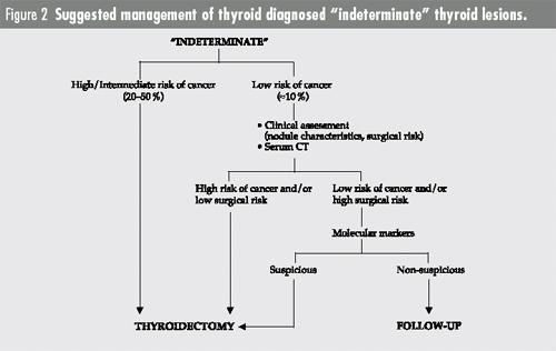
We rank “indeterminate” lesions into three classes: possibly malignant (approximately 15 % of all nodules
and with 10% cancer risk), probably malignant (approximately 1.5% of all nodules and with 20%
cancer risk), suggestive of cancer (approximately 0.5% of all nodules and with a 90% cancer risk)25.
Patients with nodules belonging to the two high-risk subgroups are addressed to immediate surgery. In
contrast, the decision to operate patients with nodules belonging to the low-risk group is taken after
balancing the individual risk of surgery against the risk of cancer after considering cytological, clinical
and echographic aspects.
-
clinical criteria
A number of studies indicate that the evaluation of clinical nodule features increases the accuracy of
FNAB and suggest that the cytological results should always be evaluated in light of clinical features8,52,53.
However, the use of clinical factors to stratify cancer risk in “indeterminate” lesions is not standardized.
Statistical methods may be used to integrate clinical and cytological information. By multiple logistic
regression we found that both age and gender significantly affect the “a priori” cancer risk of a thyroid
nodule with the risk being higher in males and at extreme ages. Similarly, also in “indeterminate”
lesions the cancer risk was higher at extreme ages8. Other authors have applied Bayesian analysis in a
group of patients with an “indeterminate” thyroid lesion to identify clinical factors predictive of cancer.
They have found that the cancer risk is higher in males than in females (43% vs.16 %), in nodules > 4
cm as compared with smaller ones (40% vs. 13 %) and in solitary nodules as compared with nodules in
a multinodular goiter (25% vs. 6%)54.
-
use of ancillary techniques
Serum calcitonin is elevated in most medullary carcinomas and should be measured in all patients with
an “indeterminate” cytological diagnosis51.
Furthermore, a variety of molecular abnormalities have been described in thyroid tumors and appear to
be relevant for tumor initiation and/or progression. On this basis, a number of studies have tried to identify
molecular markers of malignancy that could be reliably evaluated in cytological aspirates.
-
Detection of pathogenetic oncogene mutations/rearrangements
– Typical of follicular cancer: Recently, in a small series of follicular carcinomas a novel gene
rearrangement has been described involving the homeobox DNA domains of the PAX-8 gene and
the DNA binding, ligand binding, RXR dimerization and transactivation domains of PPAR-γ gene.
The resulting fusion protein has lost the activity of wild type PPAR-γ and is suspected to have a
pathogenetic role in follicular cancer55. No data are so far available with regard to the diagnostic
value of detecting PAX-8/PPAR-γ rearrangement in FNAB aspirations.
– Typical of papillary cancer: RET/PTC rearrangements are balanced inversions or translocations
that involve the intron 11 of RET and result in the translation of fusion proteins with constitutive
tyrosine kinase activity. Eleven types of RET/PTC rearrangements have been described, RET/PTC1
being the most frequent. The detection of RET/PTC by immunohistochemistry and/or RTPCR
has been proposed as a diagnostic tool to confirm the diagnosis of papillary cancer in uncertain
cases56. RET/PTC is, however, present only in 20–30% papillary carcinomas. Therefore, the sensitivity
of this procedure is low. Moreover, RET/PTC may be also present in Hashimoto thyroiditis,
Hurthle cell neoplasms and hyalinizing trabecular tumors.
– Typical of medullary cancer: Activating point mutations of the RET proto-oncogene have been
identified as a genetic pathogenetic mechanism in medullary carcinomas, either occurring in the
context of a MEN-2 syndrome or in the sporadic variant. The preoperative identification of RET
mutations in the aspirated material allows discrimination between these two possibilities, thus
influencing the therapeutic approach57.
-
Miscellaneous molecular markers
This heterogeneous group comprises a variety of molecules involved in a variety of biological functions
(e.g. cell transformation, proliferation, motility, resistance to apoptosis, differentiation) and that
maybe abnormally expressed in cancer thyroid cells. The expression of these molecules can be evidenced
by standard immunocytochemistry that could be easily included in routine thyroid FNAB process.
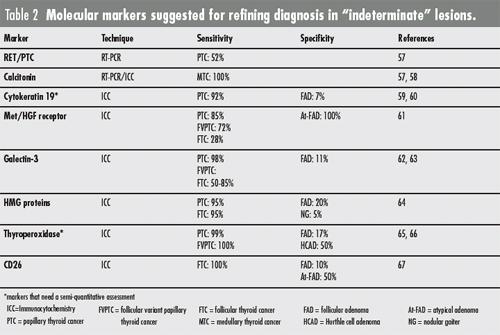
Most of these markers are able to identify papillary cancers, including FVPTC, with good accuracy58,67.
This ability could by itself justify their use since approximately 50 % of malignancies classified “indeterminate”
actually are papillary carcinomas. Widely invasive follicular carcinomas may also be positive
to these markers. However, in general, these markers have a much lower accuracy in identifying minimally
invasive follicular carcinomas, a diagnosis often problematic even at histologic examination.
Immunostaining for calcitonin has a very good accuracy in confirming the suspect of medullary cancer.
The characteristics of some of these markers are given in Table 2.
Unfortunately, the sensitivities and specificities reported are calculated from small series. Prospective studies
including a large number of patients are needed, therefore, to assess the clinical value of these markers
in refining the diagnosis of “indeterminate” thyroid lesions and altering their management. A proposed
algorithm for the management of “indeterminate” lesions is given in Fig. 2.
In aspirations from suspected cervical lymph nodes, cells of uncertain origin can be identified as epithelial
by immunocytochemistry with anti-cytokeratin antibodies and as thyroid follicular or parafollicular
cells with anti-Thyroglobulin and anti-Calcitonin antibodies, respectively. In the aspirated material the
presence of Thyroglobulin and/or Calcitonin can be also detected by conventional immunoradiometric
methods or by RT-PCR58,68.
Inadequate samples
In case of inadequate sampling simple rebiopsy is successful in approximately 50 % of cases. US-FNAB
is recommended to avoid areas of fibrosis, calcification or cystic degeneration. Repeatedly unsuccessful
FNAB does not change the “a priori” nodule cancer risk and the clinician should rely on clinical and
echographic characteristics to select the nodule for surgery
|
|


