|
| |
Thyroid FNAB smears can be classified as diagnostic or non-diagnostic. In the first group are comprised
the majority of benign and malignant thyroid lesions that can be identified with high accuracy by
standard cytological criteria. In the second group are comprised two subgroups: a) smears containing
insufficient cellular material (inadequate samples); b) smears that cannot be reliably classified either in
the benign or in the malignant group on the basis of standard cytological criteria (“indeterminate” lesions).
A detailed description of cytological features of thyroid lesions can be found elsewhere4,34,35. Herein we
will focus on basic cytological aspects that allow identification of thyroid lesions and on the characteristics
of “indeterminate” lesions, which represent a major limitation of thyroid FNAB.
Basically, the following aspects are of importance when examining a thyroid smear: a) presence of colloid;
b) cell arrangement; c) nuclear and cytoplasmic features; d) additional background features.
For instance, the presence of moderate or large amount of colloid is suggestive of nodular goiter. In
contrast, colloid is scanty or absent in neoplastic or inflammatory lesions. The evaluation of the cellular
arrangement may give indications of the histological structure. Cytoplasmic and, especially, nuclear details
may suggest a neoplastic lesion with a degree of accuracy depending on the tumor histotype. For
instance, papillary carcinoma is characterized by relatively specific nuclear features that include ground
glass cell nuclei, nuclear inclusions, groves34. Elongated “bipolar” nuclei suggest a medullary cancer36
while highly pleomorphic spindle-shaped or giant nuclei are compatible with an anaplastic cancer37. The
presence of additional features such as psammoma bodies, inflammatory cells, cell necrosis, is of major
relevance for the diagnosis of many types of lesions.
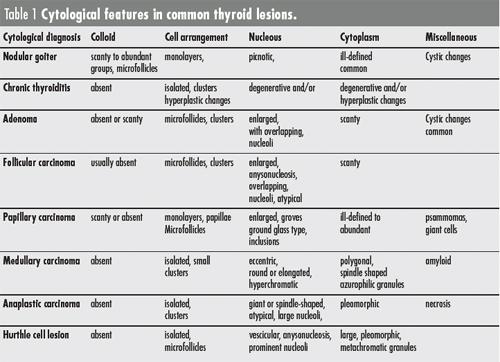
Major cytological findings of the most common thyroid lesions are summarized in Table 1.
- Diagnostic FNAB
Benign lesions, such as nodular goiter and chronic thyroiditis, and malignant lesions, such as papillary,
medullary and anaplastic carcinomas, mostly show specific cytological aspects (Table 1). They can be
diagnosed with high accuracy and are included in this group.
- Non-diagnostic FNAB
Inadequate sampling
Cytological smears should contain a good number of well preserved and well stained cells with epithelial
characteristics in order to be classified as benign. Aspiration of too few cells to reach a diagnosis is a
major limitation of FNAB. In a recent review of seven large series, the mean inadequacy rate was 17%
(range 2–21%)34. It is important to avoid making a diagnosis in the presence of a suboptimal sample, as
this is a major cause of false negatives. In order to facilitate recognition of sample adequacy, some
authors have proposed to follow quantitative criteria. Hamburger recommends that cancer cannot be
excluded if the sample does not contain at least six benign follicular cell clusters, each containing 10–15
cells, in two specimens derived from two different aspirates5. Other authors have adopted less stringent
criteria.
Variables that may affect adequacy and quality of cytologic smears have been described above.
“indeterminate” lesions
Standard thyroid FNAB is unable to reliably classify as benign or malignant a proportion of nodules ranging
15–30 %. These lesions are variably indicated as “follicular” or “suspicious” or “indeterminate” and represent
a heterogeneous group of lesions, either benign or malignant, that have overlapping cytomorphologic
aspects:
- Follicular lesions: the most common component of
the “indeterminate” group is represented by well differentiated lesions with follicular structure,
which account for 10–15% of all thyroid nodules. The large majority of these nodules are benign
tumors (follicular adenomas) or hyperplastic goiter nodules. However, approximately 1 out of
10–15 follicular lesions is malignant (well differentiated follicular carcinoma). These carcinomas differ
from adenomas because have acquired the ability to invade the tumor capsule and/or blood
vessels. On the basis of the extent of invasion follicular carcinomas are classified as minimally or
widely invasive. Cytological atypias are present in widely invasive tumors but are rare in minimally
invasive follicular carcinomas. This last category, therefore, can be only diagnosed at histopathology
after examination of multiple sections.
- Papillary carcinomas: it is increasingly recognized that, especially in areas where follicular carcinomas
are rare, papillary cancer is a significant component of malignancies diagnosed as “indeterminate”
at cytology. Most papillary carcinomas classified as “indeterminate” lesions belong to the
follicular variant (FVPTC). In FVPTC (approximately 20 % of all papillary carcinomas) a number
of cytologic features characteristic of papillary cancer, such as psammoma bodies, nuclear clearing,
grooves and pseudoinclusions, are poorly represented38,39. A small proportion of the classic
variant of papillary cancer may also scarcely present these cytological characteristics40.
- Hurthle cell lesions: Hurthle cell lesions represent approximately 2–3% of all nodules and include
neoplastic lesions (either benign or malignant) and non-neoplastic lesions, such as metaplastic
changes observed the context of Hashimoto’s thyroiditis, or nodular goiter. Hurthle cell carcinomas
represent less than 1% of all nodules and cannot reliably be differentiated from either benign
Hurthle cell tumors or non-neoplastic lesions.
- Insular carcinoma: it is an unusual thyroid tumor, representing 4–7% of all thyroid carcinomas. This
cancer is characterized by a growth pattern in nests of cells called “insulae” and by a high
aggressiveness and early spread to distant sites41. Smears are often highly cellular and poor of colloid.
However, microfollicles are rare and there is a predominance of single cells. Nuclei are predominantly
round, monomorphic and with small nucleoli. Inclusion of insular carcinoma in the
“indeterminate” group may result from the scarce signs of cellular atypia.
- Medullary carcinoma: medullary carcinoma represents approximately 5% of all thyroid carcinomas.
Aspirates are often rich of cells with characteristic cytological features (Table 1). Different morphologic
variants, however, exist42. Classification in the “indeterminate” group may result from lack of
experience with all the variants of this infrequent malignancy.
|
|


