|
Thyroglobulin (Tg) measurement is available as a marker of differentiated
thyroid carcinoma only after total thyroidectomy. Otherwise, elevations
of Tg may be caused by a variety of benign diseases including goiter, Graves'
disease and autonomously functioning thyroid nodules and do not indicate
thyroid malignancy. Suppressed Tg in patients without prior thyroidectomy
is seen under thyroid hormone medication, particularly when thyrotoxicosis
factitia is present.
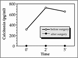
Modern immunoassay techniques have nowadays mostly replaced RIA methods, but still encounter a variety of methodological problems: Tg levels may vary among different methods used by as much as 65%. This may be improved in the future by adoption of a universal standard preparation. Major differences that are presumably related to different specificities of the anti-Tg antibodies used in various methods may still remain. Owing to lack of appropriate standardization, values that have been reported for sensitivity may not be directly comparable. As with the TSH assay the best way would be to use functional sensitivity defined as the lowest Tg- concentration that is detectable with an interassay-CV below 20%. The functional sensitivity should be below 1 ng/ml. Good assay precision is also important for measuring high serum Tg concentrations. When very high serum concentrations of Tg (>1000 ng/ml) are being measured, a high dose "hook" effect may occur. Testing the serum in a 1:10 dilution may identify this problem. Tg antibodies which are present in about 20–30% of patients with thyroid carcinoma, still constitute a major problem of interference in Tg assays. Therefore Tg measurement routinely must include validation by recovery of an external addition of Tg. Although recovery studies are useful to identify problems with interference, demonstration of intact recovery does not necessarily guarantee correctness of the value. Determination of TgAb may be of additional value in this setting. Interpretation of Tg-values should be made in appreciation of the clinical presentation of the patient. Calcitonin is available as a sensitive and specific tumor marker in the diagnosis of medullary thyroid carcinoma. Sensitivity of assay procedures has recently been improved. As for Tg, the results from different methods are not directly comparable so as to require separate standardization by each laboratory. Elevation of calcitonin almost always indicates the presence of medullary thyroid carcinoma, except for slightly elevated levels that are found in other neuroendocrine tumors, such as carcinoid, lung cancer or in pheochromocytoma and in patients with renal failure. The sensitivity of calcitonin measurement is improved by stimulation with pentagastrin. Figure 6 shows an example of a patient with MEN 2A before and after thyroidectomy for medullary thyroid carcinoma.As for thyroglobulin assays, a high dose “hook” effect may occur at very high concentrations of calcitonin. Figure 6:
|