


* Dept. of Urology, lst Medical School, Charles University, Prague
** Institute of Endocrinology, Prague
It is not without interest that while other parts of organism are slowly atrophying, prostate resumes its growth, which takes origin in so called periurethral and stromal nodules according to the McNeal's theory from 1978. So far, some growth factors have been blamed to cause this growth, but no single one is perhaps responsible. In Schreiber's review, the thesis that thyroid tissue might be originally a derivate of gastrointestinal system was submitted. Transition zone of the prostate, which is the supposed origin of the benign prostatic hyperplasia is also derivate of entodermal tissue, which is the basic embryonal tissue of gastrointestinal system. If it is true, there should be no wonder that gastointestinal polypeptides might be detected in the prostate and might be of some importance in various pathological situations. Similarly to the steroid hormones - testosteron, dihydrotestosterone or oestradiol - triiodothyronine (T3) acts directly in the cell nucleus using its receptor. Thyroxine (T4), though released in much more higher amount into the plasma, serves mainly as prohormone, or in other words, as reservoir for conversion into its active metabolit - T3.
The influence of T3 on protein metabolism is equivocal in general. It raises the catabolism of many tissues up, causing the negative nitrogen ratio. In muscles the synthesis of proteins is mainly enhanced, which may be mediated by insulin-like growth factor 1-IGFI (Límanová). In prostate, the main growth factor influencing stromal growth is basic fibroblast growth factor. Story cultured prostatic cells in androgen free medium, but in presence of growth factors. He found that stromal cells synthesize basic fibroblast growth factor (bFGF, FGF2) and keratinocyte growth factor (KGF, FGF7). Basic FGF is a potent mitogen for prostatic stroma (PS) but a weak mitogen for prostatic epithelium (PE). Begun confirmed it in his article on bFGF writing that bFGF is a potent mitogen for mesenchymal cells, including fibroblasts cultured from prostate, and it has been postulated that it plays a role in the development of benign prostatic hyperplasia. In lipid metabolism, there is reversed influence on the plasmatic level of lipoproteins, the higher thyroid hormones are, the lower levels of VLDL, LDL, or even HDL and total cholesterol are detected.
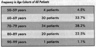
For the statement of hormones and growth factors levels, the samples of blood were centrifuged immediately and stored at -20°C in a freezing box, until the analysis was performed. The tissues were stored in a deep freezer (-80°C) and they were homogenisated in an ice salina before analysis. After centrifugation the contents of tissue hormones in a supernatant were determinated in the same way as the hormones in serum. The hormones of the thyroid gland in serum or tissue in patients with BPH or prostatic cancer were performed using: thyroxine (RIA, code RI 241), triiodothyronine (RIA, code RI 271 ), the pituitary hormone TSH by ILMA (code LI 022), all from Immunotech a.s., Prague, Czech Republic, and basic fibroblast growth factor (bFGF) in tissue by ELISA, code RPN 2158, Amersham Int. U.K. The volume of the prostate was measured by transabdominal way using ultrasound probe 3.5 MHz. The number of patients with BPH and PC in single decades were as follows - Fig. 2
|
|
|
Considering the relations between sonographically measured volume and TSH, TSHT, T3 and T4, no significant relation at least at the level p < 0.05 was found despite the fact, that in case of sono / T3 in the whole group of BPH and PC only a "close shave" prevented the significant relation (r = 0.14, y = 7.07x + 33.07, N = 79) and in the BPH subgroup being expressed even closer, though insignificant (r = 0.16, y = 8.56x + 32.41, N=65).
As far as the relation between TSH, TSHT, T3 and T4 was considered (TSH / T3 and TSH / T4 were excluded due to the well known reasons), the strongly positive correlation was found between TSH and TSHT (r = 0.46, y =144.02x - 2.35). Further interesting correlation - the negative one - was found between TSHT and T4 for both: BPH + PC and even stronger for BPH alone (r = -0.22, y = -4.39x + 519.60, N = 78, and r = - 0.30, y = -6.26x + 656.96, N = 64 respectively).
Taking bFGF into account, then only positive correlation has been found between basic fibroblast growth factor and the level of thyroxine in the plasma (r = 0.25, y = 0.14x + 4.08, N = 79). The results are demonstrated in Fig. 3
Contact address:
Dr. L. Safarík
Dept. of. Urology
1st Medical School
Charles University Prague
Ke Karlovu 6, Praha 2
[2]Guess HA: Epidemiology and Natural Historv of Benign Prostatic Hyperplasia. Urol Clin N Am. May 1955. 22: 247-261.
[3] Hirsch-Marie H, Delafontaine D: Seminal pepsinogen: quantification and corelation with the spermiogram. Internat J Androl 1978. 1: 397-404.
[4] Límanová Z, Nemec J, Zamrazil V: Nemoci stítné zIázy, Galén. 1995
[5] McNeal JA: Origin and Evaluation ot Benign Prostatic Enlargement, Invest Urol 1978. No. 4, 15: 340-345. [6] Mebust WK, Holtgrewe HL, Cockett ATK. Peters, amd Writing Committee: Transurethral prostatectomy: immediate and postoperative complications. A cooperative study of 13 participating institutions evaluating 3,885 patients. J Urol 1989. 141: 243-7.
[7] Reese JH, McNeal JE. Redwine EA. Samloft LM. Stamey TA: Differential distribution of pepsinogen II between the zones of the human prostate and seminal vesicle. J Urol 1986, 136: 1148-1152.
[8] Schreiber V: Enigmas of the Thyroid Gland. The Thyroid Gland. 1996, 1:3-6.
[9] Story-MT: Positive and negative modulators of prostate cell proliferation Biomedicine-and-Pharmacotherapy. 1994 48/SUPPL. 1, 35S-41S.
[10] Walsh PC: The Prostate. Johns Hopkins Book, 1995.