|
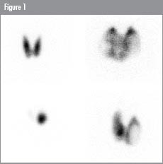
Benign thyroid diseases The most common indications for thyroid scintigraphy are, in decreasing frequency: hyperthyroidism, euthyroid multinodular goiter, and hypothyroidism. Imaging for all three indications can be performed with either 123I, 131I, or 99mTc-pertechnetate. The advantage of using isotopes of iodine are that the amount of thyroidal uptake of radioactivity of the images can be used to compute the amount of 131I that is to be administered for therapy of hyperthyroidism or goiter. With 99mTc this cannot be done reliably because of the rapid wash-out of the radioactivity and because of the relatively low target-to-background ratios. If logistically possible, 123I should be preferred over 131I, because of the lower patient radiation burden, and also if diagnostic scanning is followed by treatment with 131I, because of the absence of so-called “stunning”, i.e. lower retention of 131I at the time of treatment due to a radiation effect by the beta-emission of the diagnostic dose of 131I. In case of thyrotoxicosis, 4 distinct patterns of radioiodine uptake in the thyroid can be encountered:
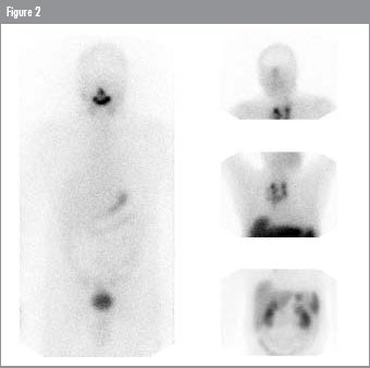
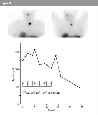
Thyroid malignancy It makes sense to distinguish between imaging methods used for investigation of possible thyroid malignancy and those that can be applied to localize metastatic disease. Radioiodine imaging of the thyroid is certainly of great value in patients with nodular thyroid disease as increased uptake of the isotope in the nodule as compared to the surrounding tissue renders malignancy less likely. However it may be questioned whether there is a role for any nuclear medicine imaging technique for diagnosis of malignancy: fine needle biopsy cytology or pathology of removed tissue will always be necessary to reach the diagnosis. Apart from this, all currently available radiopharmaceuticals have a relatively low specificity for detecting malignancy: only 20% of “cold” nodules on radioiodine or pertechnetate scans are malignant2; medullary cancer may be missed with somatostatin receptor imaging (SRI)3; both benign and malignant thyroid nodules may accumulate 18Fluoro-Deoxy-Glucose (18FDG)4; other imaging agents like 201Thallium (201Tl), 99mTc-(V)-dimercapto succinic acid (DMSA), and 99mTc-methoxyisobutylisonitrile (MIBI) all lack specificity5,6. It can therefore be concluded that nuclear medicine has no “magic bullet” to identify malignancy in thyroid nodules. Quite different is the situation in the follow-up of patients who have been operated for thyroid cancer. In patients with differentiated thyroid carcinoma (DTC; papillary and follicular carcinoma), surgery is followed by ablative therapy with 131I, and further follow-up imaging, guided by serum thyroglobulin (Tg) concentrations, whether during TSH suppression or just serum TSH elevation, consists of radioiodine scanning as long as the tumour is radioiodineavid. The reason for this policy is twofold: 1. If a limited number of lesions is identified, they can be removed by (probe-guided) surgery; 2. If further surgery is precluded, diagnostic scanning can be followed by high dose 131I therapy. In patients with detectable serum Tg concentrations but negative post-therapy 131I scans, there are several nuclear medicine techniques that may demonstrate tumour sites. 201Tl whole-body scintigraphy can be used to identify metastatic sites of DTC. After concentration of 201Tl via the ATPase-mediated sodium-potassium pump, the tumour uptake is dependent on, amongst others, vascularity and cellularity of the tumours, and also prolonged retention in the tumours plays a role7,8. The sensitivity in detecting metastatic sites is 60–90% and does not require (endogenous or exogenous) TSH stimulation9–12. 201Tl scanning is not specific for thyroid carcinoma, however, as other tumours also concentrate the radiopharmaceutical. 99mTc-MIBI is another radiopharmaceutical that can be used to image DTC. The exact mechanism of 99mTc-MIBI tumour uptake is not known, but the uptake is related to the intracellular mitochondrial markdensity. It has been used in thyroid carcinoma, with high sensitivity13,14. 99mTc-(V)-DMSA resembles the phosphate ion and readily accumulates in calcifying tumours like medullary thyroid carcinoma (MTC). The sensitivity in patients with MTC is high15. False negative findings, however, are more frequent in patients with minimally increased serum tumour markers. Over the last years an increasing amount of papers have been published that stress the importance of 18FDG Positron Emission Tomography (PET) scanning in patients with recurrent thyroid carcinoma. Because most tumours have an increased metabolism and also an increased number of glucose transporter proteins, they can be imaged with 18FDG, a glucose mimic that remains trapped in the tumour cells. Due to the fact that many tumours, but also infections, show increased glucose metabolism, the specificity of 18FDG PET imaging for recurrent or metastasized thyroid carcinoma may be low, but sensitivity is high. Several groups have reported sensitivities of 80–95% for both MTC and DTC16–19. A new development that is expected to gain importance in the clinical decision making in patients with thyroid cancer is combined CT-PET camera imaging, which has a high image resolution and combines functional with anatomical information. As with other imaging techniques, the value of 18FDG PET is greatest in patients with DTC who have elevated serum Tg concentrations but in whom no tumour uptake is found with 131I. The results of this type of investigation in terms of tumour to background contrast and tumour detection is better after TSH stimulation, whether by thyroid hormone withdrawal or stimulation with recombinant TSH20,21. Somatostatin Receptor Imaging (SRI) can be used to demonstrate MTC and DTC. Somatostatin receptors are present on a variety of neuroendocrine tumors, and these can be visualized with the radiolabelled somatostatin analogue [111In-DTPA0] octreotide. In patients with medullary thyroid carcinoma (MTC) the sensitivity of SRI to detect tumour localizations is 50–70%3,22. In a series of 17 patients with MTC whom we studied3, the ratio of serum calcitonin over carcinoembryonic antigen (CEA) levels was significantly higher in patients in whom SRI was successfully applied. This may imply that somatostatin receptors can be detected in vivo on the more differentiated forms of MTC. Also, SRI is more frequently positive in patients with high serum tumour markers and large tumours22, and seems therefore less suitable to demonstrate microscopic disease22,23. Although papillary, follicular and anaplastic thyroid cancers, and also Hürthle-cell carcinomas, do not belong to the group of classical neuro-endocrine tumours, the majority of patients with these cancers show uptake of radiolabeled octreotide during SRI24,25 (Figure 2). Also here, it is not necessary to use TSH stimulation to perform SRI26. Interestingly, also differentiated thyroid cancers that do not take up radioactive iodine, may show radiolabeled octreotide accumulation24. In some patients, this could open new therapeutic options: operation, if the number of observed lesions is limited, or, alternatively, Peptide Receptor Radionuclide Therapy (PRRT; see below) if the uptake is sufficient. In patients with somatostatin receptor-positive pathology, SRI is useful if it can localize otherwise undetectable disease, or if it can be used for treatment selection (usually the choice between symptomatic treatment with somatostatin analogues or other medical treatment). In patients with known metastatic disease, however, in whom little or no treatment alternatives are available, SRI has a limited role. This situation changes if imaging has a sequel in PRRT. Because of the promising results in patients with neuroendocrine tumours of treatment with Auger electrons or beta-particle emitting somatostatin analogues27–30, we expect that SRI will be increasingly used to select patients for such therapy (Figure 3). Beta-particle emitting octreotide analogues used for PRRT are [90Y-DOTA0,Tyr3]octreotide and [177Lu-DOTA0,Tyr3]octreotate, which have a 3 to 4-fold higher uptake than [111In-DTPA0]octreotide in most neuroendocrine tumors31. PRRT with these analogues leads to an anti-tumor effect in about twothirds of the patients with neuro-endocrine tumors. It should be taken into account however, that in most patients with MTC the uptake of radioactivity in the tumour sites is low if compared with other neuroendocrine tumours like carcinoids and endocrine pancreatic tumours. Also, in our experience, in most patients with DTC the tumour uptake after [177Lu-DOTA0]octreotate is not higher than after [111In-DTPA0]octreotide. This is most likely due to the fact that not the somatostatin receptor-subtype 2 is overexpressed in these tumours, but other subtypes, in agreement with an in vitro study using RT-PCR on human thyroid carcinoma cell lines and demonstrating a predominance of mRNA for somatostatin receptor subtype 3 and 5, but only very low amounts of mRNA for subtype 232. [90Y-DOTA0,Tyr3]octreotide has a higher affinity for the somatostatin receptor subtype 5 than [177Lu-DOTA0,Tyr3]octreotate, but its importance for PRRT of DTC has to be shown. It is to be expected that in the near future somatostatin analogues will be introduced for SRI and PRRT that show higher affinity for the somatostatin receptor subtype 3 and 5, in addition to a high affinity for the subtype 2. This will open a new area for the staging and treatment of DTC. Cholecystokinin-B (CCK-B) receptors, which bind both CCK and gastrin, have been demonstrated in human medullary thyroid carcinoma33. The presence of this receptor is probably the molecular basis for the well-established pentagastrin stimulation of serum calcitonin (CT) to diagnose otherwise undetectable MTC. In a study using in vivo scintigraphy with an 111In-labelled CCK analogue in 7 patients with biochemical or radiological evidence of MTC, presumed tumour sites were demonstrated in 2 patients34. The uptake was however low (Figure 4). Behr and Béhé, using a different analogue, were able to demonstrate tumour sites in all of 23 patients with known tumour localizations of MTC, as well as in 20 of 22 patients with occult disease35. Subsequently, they also treated MTC patients with 90Y-labelled gastrin analogue, with promising results in terms of tumour growth control. Therapy related side-effects like myelodysplastic syndrome and nephrotoxicity, however, are still major concerns to be solved.
|