|
With the advent of newborn screening clinical studies of affected infants and
advances in molecular medicine have greatly expanded our understanding and nosology
of CH. The average prevalence varies from 1: 3500 to 1: 4000 neonates.27,28 The
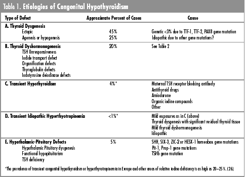
types of defects associated with CH and the known causes are summarized in table 1.
Approximately 70 % of cases are due to thyroid dysgenesis, either ectopia or
hypogenesis-agenesis.24,28,30 Relative prevalence as high as 85 % has been
reported.23 Two thirds of the infants with thyroid dysgenesis have some residual
thyroid tissue.28,29 Thyroid dysgenesis is more prevalent in female infants
(F/M–2/1), is less prevalent in black (1: 32000) and more prevalent in Hispanic
infants (1:2000).27,29,31 Approximately 2% of cases are familial and mutations in
the homeobox genes TTF-1, TTF-2, and PAX-8 have been described in such
patients.30,31 The prevalence of congenital anomalies in infants with congenital
hypothyroidism is increased (7–20% vs. 3.2 % in the general population);
defects include congenital heart disease, hip dislocation, cleft palate, Down
syndrome, trisomy 18, and dysmorphic syndromes (Beckwith, Sotos).22,32 Congenital
malformations are more prevalent in infants with transient CH than in those with
permanent disease presumably due to increased iodine antiseptic exposure, birth
asphyxia, respiratory distress or other morbidity associated with the malformation
in the neonatal period.32 The prevalence of CH is estimated to be 1 in 41 cases of
Down syndrome, a prevalence 100 times more frequent than in the general population.33
In the familial cases there are congenital malformations associated with the
homeobox gene mutations. In the much larger number of non-familial cases the etiology
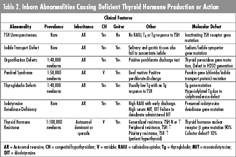
remains obscure. Transient congenital hypothyroidism (low serum T4 with elevated TSH) has been observed in association with iodine or organic iodinated compound exposure of the mother or infant, maternal antithyroid drugs or other therapeutic agents (amiodarone, anti-asthmatics), and maternal anti-TSH receptor blocking antibodies.22,23,34,35 The presence of thyroid gland enlargement in the neonatal period is evidence of drug or goitrogen-induced hypothyroidism, but most infants with transient hypothyroidism have normal thyroid glands by palpation and ultrasound. Radioiodine uptake will be inhibited by iodine or TSH receptor antibody and scan results in such infants will be variable. Transient hypothyroidism is of short duration (1–2 weeks) in the case of drug exposure, but will be of longer duration (1–4 months) if related to maternal TSH receptor blocking antibody, the half-life of which approximates 2 weeks.35 Transient hypothyroidism should be treated if severe and/or persists beyond 1–2 weeks. Transient hyperthyrotropinemia with a normal T4 concentration is an uncommon disorder. The usual cause is a transient and mild impairment of thyroid hormone secretory capacity due to thyroid dysgenesis, thyroid dyshormonogenesis, or TSH receptorblocking antibody.36 Idiopathic hyperthyrotropinemia is a rare disorder described in Japan where it is detected in 1 in 18,000 newborns.37 In a recent report of 16 cases followed for 2–7 years the elevated serum TSH levels at 2–7 weeks ranged from 17 to 77 mU/L with normal T4, T3 and free T4 concentrations; basal metabolic rates and thyroid radioiodine uptake were also normal. TSH responses to TRH were increased and TSH receptor antibody was not detected. Serum TSH normalized spontaneously in 11 of 16 infants within 6 months but the increased response to TSH persisted for 3–7 years. All had a normal T3 response to TSH excluding a hormone synthetic defect. However, small diffuse goiters developed in 3 of 16 cases by 5–7 years. The etiology has not been defined. Defects associated with Thyroid Dyshormonogenesis are summarized in table 2. The function of the thyroid gland is to concentrate iodide from the blood and return it to peripheral tissues in a hormonally active form. The major substrates for thyroid hormone synthesis are iodide and tyrosine. The process of thyroid hormone biosynthesis is stimulated by TSH binding to the follicular cell TSH receptor, intracellular transduction of the signal to G proteins and production of cAMP (Figure 1). Processes stimulated by cAMP include: cell membrane iodide transport, thyroglobulin synthesis, oxidation and organification of trapped iodide at the colloid-cell membrane interface, activation of colloid endocytosis and intracellular phagolysosome formation, and hydrolysis of thyroglobulin (Tg) to release monoiodotyrosine and diiodotyrosine, T4 and T3. The iodotyrosines are largely deiodinated within the thyroid cell; T4 and T3 are released into the circulation. Significant amounts of Tg also escape from the gland, predominantly via the thyroid lymphatic system. Thyroid dyshormonogenesis usually is transmitted as an autosomal recessive trait. Except for the familial incidence and tendency for affected individuals to develop goiter, the clinical manifestations of CH due to a biochemical defect are similar to those in infants with thyroid dysgenesis. Thyroid enlargement may be conmanifest at birth, but in many patients development of the goiter is delayed. There are several recent reviews of these disorders.24,38-41 Laboratory analyses to characterize the genetic defects in children with thyroid dyshormonogenesis are not yet generally available. Hypothalamic-Pituitary hypothyroidism is a significant cause of CH occurring in 1:20,000 to 1:50,000 newborns, but usually is not detected in newborn programs screening for increased serum TSH levels.23 Disorders associated with hypothalamic-pituitary TSH deficiency include anencephaly, holoprosencephaly, septo-optic dysplasia, median facial dysmorphic syndromes, pituitary hypoplasia/aplasia, and single or multiple pituitary hormone deficiencies. The pathogenesis of most of these disorders remains obscure, but mutations of three homeobox genes, sonic hedgehog (SHH), SIX-3 and ZIC-2 have been described in infants with holoprosencephaly.42-44 HESX-1 homeobox gene mutations have been associated with septo-optic dysplasia involving midline brain defects and pituitary hypoplasia.45 Homeodomain genes involved in hypothalamic development in the rodent include SF-1, LHX-3 and LHX-4.46 Prop-1 and Pit-1 genes are terminal factors in the homeobox gene cascade programming development and function of pituitary cells producing growth hormone, prolactin, TSHß, and GHRH receptors. Mutations of Prop-1 or Pit-1 have been described in patients with familial hypopituitarism.47 Mutations in the TSHß gene also have been reported in rare patients with TSH deficiency.48
Figure 1 |