|
In order to understand the
effect of thyroid hormones on bone it is important to describe in
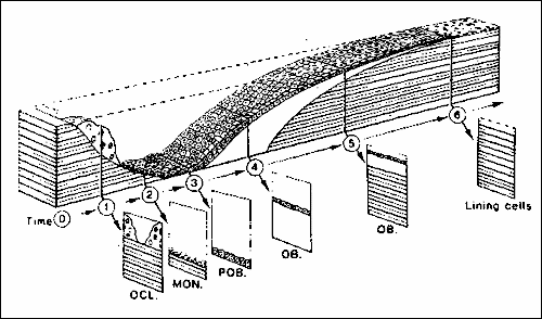
some detail the normal process of bone turnover. In adults, there
is a continuous process of both bone resorption and bone
formation which are tightly coupled, so that the net activity of
bone resorbing cells equals the net activity of bone forming
cells. This process of bone resorption followed by bone formation
is called remodelling (see also figure page 4). It provides a
mechanism for self repair and adaptation to stress. Bone
remodelling is a cyclic process, occurring dyssynchronously at
thousands of sites in the human skeleton at all times. Because of
its high surface to volume ratio, many more remodelling sites are
to be found per unit volume of trabecular than cortical bone.
Therefore, disorders of remodelling become earlier manifest in
trabecular than in cortical bone.
Remodelling starts with activation, the attraction of bone resorbing cells (osteoclasts) to a locus on the bone surface. Activation frequency describes the frequency with which activation events occur on bone surfaces, in other words "how often a given site on the bone surface undergoes resorption and subsequent formation". The osteoclasts create a resorption cavity, a process which takes 4 to 12 days. The depth of the cavity is approximately 40–60 µm. After a reversal phase, osteoblasts start to invade the area and begin bone formation. First an osteoid matrix is formed, which is later mineralised. The total remodelling sequence takes approximately 200 days. Normally, the amount of bone formed, equals the amount which was first resorbed. The end product of the remodelling sequence is the bone structural unit (BSU).
Thyroid hormones exert their effect on the activation frequency such that higher levels of thyroid hormone lead to a higher activation frequency. In other words, there is an increase in the total number of activated BSUs. In addition, both the resorption and the formation phase of the remodelling sequence are shortened. In thyrotoxicosis, the surface area of unmineralised matrix (osteoid) is increased. Although both osteoblast and osteoclast activity are increased by elevated levels of thyroid hormone, the net result is loss of bone mass. The final resorption depth is normal, but the amount of bone formed is decreased, leading to a negative balance of approximately 10 µm per remodelling cycle. Apparently, the shortening of the formation phase predominates. The latter, together with the increase in total ongoing BSUs lead to an increase in the remodelling space (i.e. the space corresponding to the amount of bone resorbed by osteoclasts but not yet reformed by osteoblasts). When thyroid hormone levels are decreased, the activation frequency is reduced, and the resorption and formation phases are prolonged. In states of hypothyroidism, the resorption depth is reduced, but the amount of bone that is formed is near normal, despite a reduction in osteoblastic activity. This leads to a positive balance of approximately 15 µm per remodelling cycle. Although thyroid hormone stimulates both osteoblasts and osteoclasts, stimulation of the latter appears to be indirect. The exact mechanism by which thyroid hormones increase osteoclast activity remains unknown, but in vitro experiments show that the presence of (activated) osteoblasts is a prerequisite for stimulation of osteoclasts. Unlike osteoclasts, osteoblasts possess nuclear receptors for T3, based on biochemical binding studies and detectable mRNA levels for thyroid hormone receptors. How stimulated osteoblast increase osteoclast activity is still unsolved. It has been suggested that the effect is mediated by a soluble factor, termed osteoclast resorption-stimulating activity, which appears to be associated with the osteoblast cell surface or the extracellular matrix. During the various stages of bone turnover, markers of this process are released are released. These biomarkers provide the possibility of assessment of bone resorption and formation. The rate of formation and resorption of bone can be assessed by measuring enzymatic activity related to bone-forming (alkaline phosphatase) or resorbing (acid phosphatase) cells. An alternative is to measure bone matrix components released into the circulation, either after their secretion by osteoblasts (osteocalcin, procollagen type I) or after their release (as breakdown products) during osteoclastic bone resorption (urinary hydroxyproline, pyridinoline crosslinks), see also the table. In hyperthyroidism, as a result of coupling, both markers of bone formation and of bone resorption are increased. In fact, there is a significant correlation between serum alkaline phosphatase and thyroid function. On the other hand, most markers of bone turnover are decreased in patients with hypothyroidism. As a result of increased bone resorption, serum calcium is also slightly higher in subjects with hyperthyroidism, although serum levels above the upper level of normal are rare. This increase in serum calcium will lead to suppression of PTH-secretion and to a decreased formation of 1,25-dihydroxyvitamine D. Subsequently, calcium excretion by the kidney is enhanced and calcium resorption by the intestines is decreased. Tubular reabsorption of phosphate is increased. The increased renal calcium loss and the decreased intestinal calcium resorption prevent further hypercalcaemia, at the expense of a negative calcium balance. In hypothyroidism the opposite is seen, although abnormalities in serum calcium are even more uncommon than in hyperthyroidism. Table: Markers of bone turnover
|