|
In patients with autoimmune thyroid disease, an increased glycosaminoglycan
(GAG) production is observed in the orbital space, in the pretibial region,
and in cardiac valves (112). GAG are long, unbranched polysaccharide chains
composed of repeated disaccharide units. After synthesis in fibroblasts,
most of the GAG are released into the extracellular matrix. Being hydrophilic,
they attract large amounts of water, thereby forming hydrated gels even
at very low concentrations (113,114). The augmented secretion and accumulation
of GAG in the cardiac valve leads to thickening of the leaflets. Additional
disturbance of collagen synthesis causes prolapse of redundant and thickened
(> 5 mm) mitral valves, also called myxomatous mitral valve prolapse (MVP)
into the left atrium. An increased prevalence of MVP has been reported
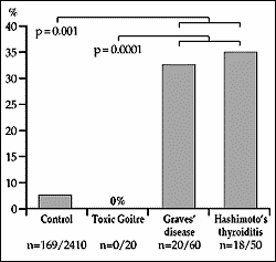
in autoimmune thyroid disease (115,116). In our clinic, the prevalence
of myxomatous MVP was investigated in patients with Graves' disease and
Hashimoto's thyroiditis, in subjects with toxic goitre, as well as in euthyroid
controls (fig.5). Myxomatous MVP was present in 36 and 33% of the patients
with thyroiditis and Graves' disease, respectively, but in none of the
toxic goitre group (117-119). In patients with thyroiditis, mitral regurgitation
was observed in 5 cases (28%), whereas prolapse of the aortic and tricuspid
valves was seen in 4 (22%) and in 1 (6%), respectively. Thyroid function
did not influence the incidence and intensity of the prolapse, thus valvar
prolapse was not attributable to myocardial dysfunction caused by a direct
effect of thyroid hormones.
Figure 5:
A potential link between autoimmune thyroid disease and myxomatous cardiac valves is suggested by histochemical studies of tissues affected directly in these disorders (120). An essential pathologic characteristic of MVP is a significant increase in the amount of GAG normally found in the superficial zone of connective tissue on the ventricular surface of the floppy mitral valve cusp. Mucinous changes extending into the elongated chordae tendinae further increasing the redundancy required for interchordal hooding or prolapse, have been seen. Like the GAG infiltrate of Graves' ophthalmopathy, these histochemical changes in the cardiac valves may be immunologically mediated. Proof of a common autoimmune mechanism requires immunohistologic studies of cardiac valves, thyroid, and orbital tissue. Given the cardiac (mitral regurgitation, endocarditis, thromboembolism, arrhythmic sudden death) and neurologic (cerebral embolic event) complications of the pathologic forms of MVP (121,122), physicians should look carefully for myxomatous involvement of the mitral valve in patients with autoimmune thyroid diseases. Patients should be monitored and prophylactic antibiotic treatment recommended when appropriate. |