|
An abnormal left ventricular function has been observed
during exercise in H, independent of ß-adrenoceptor activation, suggesting
a reversible functional cardiomyopathy due to a direct effect on the myocardium
of an excess in circulating T3 (98). Left ventricular ejection
fraction was also decreased in patients with hypo at rest and after exercise
and increased after T4 therapy (99). Furthermore, H has been
implicated as a primary cause of decreased exercise tolerance (100,101).
Stress echocardiography and cardiopulmonary exercise testing are actually
considered as reliable techniques for the evaluation and follow-up of cardiorespiratory
function (102,103). Using these non-invasive methods, 60 patients with
H were examined and controlled 6 months later in euthyroidism. In patients
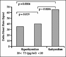
with H, heart rate showed a markedly lower increase between rest and maximal
exercise compared to the euthyroid state (fig. 3), whereas delta heart
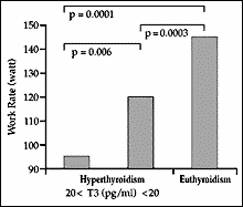
rate was strongly reduced in patients over 50 yr. Maximal work rate was
markedly reduced in severe H (fig. 4). Stress echocardiography revealed
blunted responses of ejection fraction and cardiac output during exercise
in H, and these changes were more pronounced in older patients. Compared
to controls, forced vital capacity and forced expiratory ventilation during
the first second were decreased, significantly more in older patients.
During testing, markedly decreased oxygen pulse at the ventilatory anaerobic
threshold and at maximal exercise was observed in H. Thus, especially in
older patients with H, marked but reversible alterations of cardiac exercise
capacity and cardiorespiratory function were observed. This was demonstrated
best by the reduced oxygen pulse at the anaerobic threshold, an objective
parameter for effective cardiopulmonary exercise capacity. Furthermore,
the impaired chronotropic and inotropic response during exercise might
be the primary limiting factor of reduced work capacity in patients with
H.
Figure 3:
Figure 4:
|