|
Thyroid hormones exert marked influences on electrical impulse generation
(chronotropic effect) and conduction (dromotropic effect). T3
increases the diastolic depolarisation rate, and decreases the action potential
duration, the refractionary period of the atrial myocardium as well as
the atrial/ventricular nodel refraction period. In a double-heart model,
T4 increased similarly the heart rate of the enervated infrarenal
and the innervated in situ hearts (38). In another study (58), the heart
rate in both thyroidectomized and hypophysectomized rats decreased similarly
and proportionally to T3 levels. In the same study, the effects
of thyroidectomy and chemical sympathectomy on the heart were compared.
The group with thyroidectomy had a significantly slower heart rate than
the group with sympathectomy. However, both groups responded with a similar
increase in heart rate after treatment with T3, suggesting a
direct chronotropic effect of T3. In vitro studies found that
T3 decrease the duration of the repolarization phase of the
membrane action potential and increase the rate of the diastolic depolarisation
and therefore the rate of contraction (58-60). Furthermore, studies using
an isolated heart model found that hearts from animals with experimental
H show increased heart rates and shorter mean effective refractory periods
than hearts from euthyroid animals (61). The mechanism by which T3
induce the electrophysiological changes is related in part to its effects
on sodium pump density and enhancement of Na+ and K+ permeability (62).
In humans, chronotropic effects of thyroid hormones have been assessed
using 24- hour ECG recordings. Patients with H show an increase in heart
rate throughout sleeping and wake hours (63), whereas in hypo patients
a decrease in basal, average, and maximal heart rates was found even though
most of them were not bradycardic at rest. After treatment, heart rates
in both groups returned to normal (64).
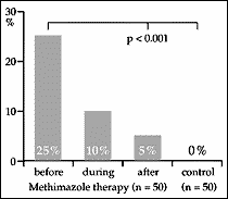
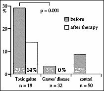
Heart rate and the arrhythmia profile were analysed in patients with H, before, during and after an antithyroid therapy (63). The number of patients with supraventricular premature complexes was elevated compared to controls (88% vs. 30%, p<0.001), and decreased markedly after therapy (fig. 1). Prevalence of supraventricular arrhythmia was age-related before as well as during antithyroid treatment, whereas ventricular arrhythmias were present in 29% of the patients with toxic goitre (median age 59yr) in contrast to 3% only of the cases with Graves' disease (37yr, fig. 2). In another study (65), the efficacy of the calcium channel-blocking drug Diltiazem in lowering the incidence of arrhythmias was evaluated. Heart rate and the number of ventricular extrasystoles significantly decreased but returned to baseline values after Diltiazem was discontinued. In a further paper (66), circadian rhythm of heart rate was maintained in H, although heart rate variability was significantly increased, supporting the view that normal adrenergic responsiveness persists in H. The prevalence of premature contractions was not different before and after therapy, although premature atrial contractions were more prevalent during the middle of the day. In conclusion, ventricular arrhythmias are rare in H patients without cardiac disease. The prevalence of ventricular arrhythmias remains essentially unchanged during antithyroid therapy and is comparable to that of a normal population. Antiarrhythmic therapy is definitely not necessary in these patients. Figure 1:
Figure 2:
In patients with hypo, only atrioventricular blocks, sinus bradycardia, and rare episodes of "torsade de pointes" have been reported. In an animal model of ventricular fibrillation, hypo increased the fibrillatory threshold of the ventricles (67). In humans, the prolongation of the QTc interval encountered in hypo is similar to that seen in euthyroid patients on class 3 antiarrhytmic agents. Finally, in patients with hypo, T4 replacement therapy did not increase significantly the frequency of benign atrial or ventricular premature beats (64,68). |