|
| |
Technically, ultrasonography of the thyroid requires the use of a linear
sector scanner in a B-Mode with a 5 to 7,5 mHz frequency. In addition a
3.5 to 5 mHz transducer may be advantageous for a particularly large thyroid
gland and substernally located parts of the thyroid. Focal lesions exceeding
a size of three mm in diameter should be reliably identified. Color Doppler
sonography may aid in visualising vascularisation of the entire gland and
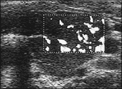
of thyroid nodules. An example of diffuse hypervascularisation in a patient
with active Graves' disease is depicted in Figure 7. Hypervascularisation
within a thyroid nodule may indicate malignancy. However, further studies
are needed to define the role of this technique in the diagnostic workup
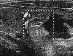
of thyroid nodules. Figure 8 shows Color doppler imaging of an autonomously
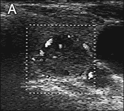
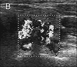
functioning thyroid nodules and Figure 9 of a medullary thyroid cancer.
The documentation of a thyroid ultrasonography examination
should include:
- calculation of thyroid volume
- echo pattern of the thyroid gland (normal / hypoechoic; homogeneous/inhomogeneous)
- localization and size in three dimensions of focal lesions and nodules
- structure (solid/liquid/calcified) and echoic pattern (normal/hypoechoic/hyperechoic) of focal lesions and nodules
- neighbouring organs such as lymph nodes and the parathyroid glands.
| Figure 7: |
Figure 8:
|
|
|
Color doppler scan of the thyroid of a patient with active
Graves' disease showing diffuse hypervascularisation.
|
Color doppler scan of the thyroid of a patient with an autonomously
functioning thyroid nodule showing the typical perinodal hypervascularisation.
|
Figure 9:
|
|
|
| Color doppler scan of the thyroid of a patient with medullary carcinoma.
In panel A a weak intranodal flow is shown which is strongly enhanced
after intraveneous application of a galactose-based sonographic contrast
agent (panel B).
|
|
|


