 Figure 3:
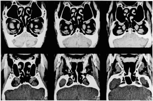
Figure 3:
Computed tomography of the orbit in a patient with dysthyroid optic neuropathy. Successive
coronal sections show marked enlargement of all four rectus muscles and apical crowding; note
effacement of the perineural fat circumventing the optic nerve in the apex of the orbit.