 Figure 9:
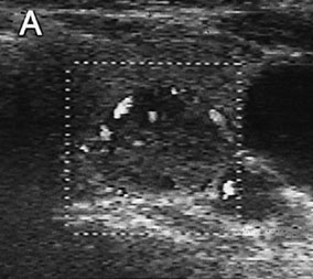
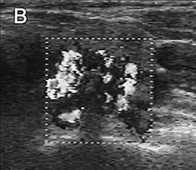
Figure 9:
Color doppler scan of the thyroid of a patient with medullary carcinoma.
In panel A a weak intranodal flow is shown which is strongly enhanced
after intraveneous application of a galactose-based sonographic contrast
agent (panel B).